Label the Photomicrograph Based on the Hints Provided
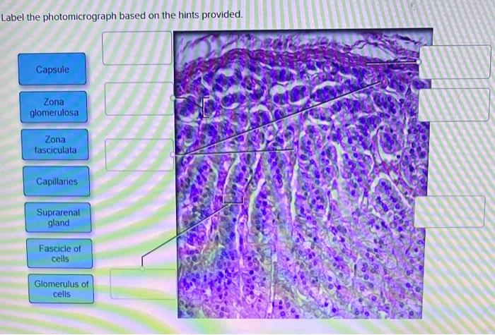
Zona glomerulosa Suprarenal gland Medullary vein Cortex Zona reticulars Modula Zona tasciculata Capsule Of Reset Zoom Prev 6 of 50H Next TV MacBook Pro 2 A 1 赴 3 4 96 5 7 2 09 8 9 o W E R T т Y Y C 0 Р 4 S D F G H J к V Z х C V B N M. Pancreas Label the photomicrograph based on the hints provided.

Endocrine Connect Lab Review Week 4 Youtube
Introduced from outside the body 2.
. View the full answer. Label The Photomicrograph Based On The Hints Provided - Solved Lab 5 Exercise 5 13 In The Photomicrograph Below Of Compact Bone Tissue Find And Label The Indicated Structures Osteer Lamella Lacuna Osteo Course Hero. Suprarenal gland Label each of the following histology slides by dragging the histology slide of the gland under the correct name.
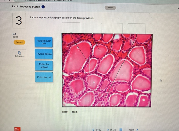
Label the photomicrograph based on the hints provided. Label the photomicrograph based on the hints provided. Molecules produced by the body 4.
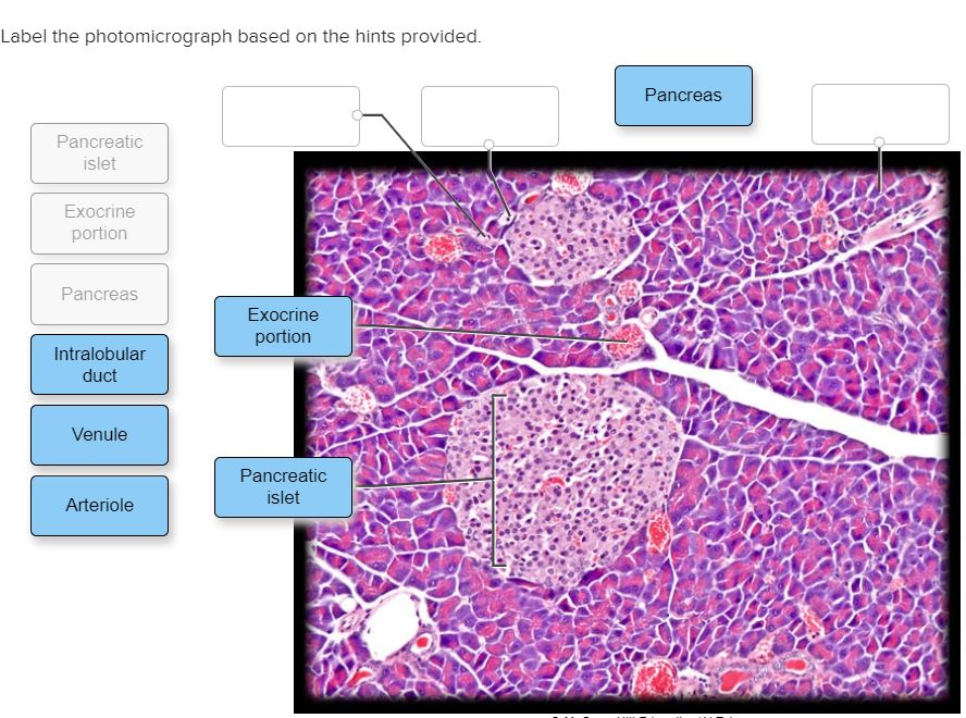
Endocrine Lab Worksheet Label the photomicrograph based on the hints provided Capillary 025 points Exocrine portion Pancreas Print Pancreatic islet References Feb 28 2022 0859 AM 1 Approved Answer. Label the structures in the photomicrograph based on the hints provided. Label the photomicrograph based on the hints provided.
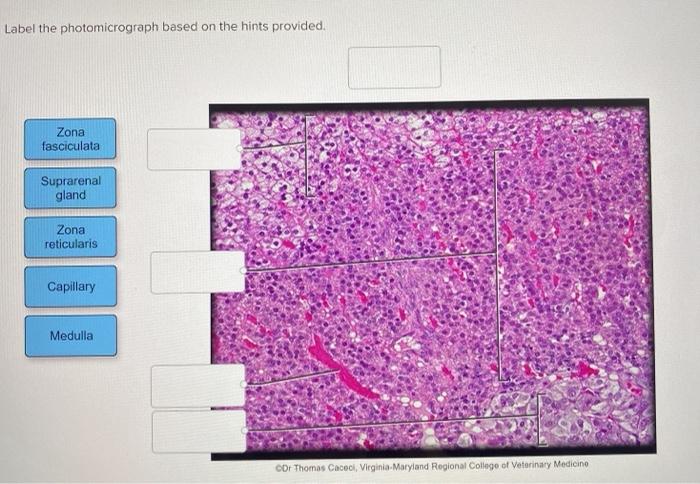
Medulla capillary zona fasciculara suprarenal gland zona reticularis. Classify each of the following parts of the nephron into the correct category based on whether it can only be found in the cortex or if it can be found in the medulla andor cortex of the kidney. Estimates range from 14 to 89 based on autopsy series.
The chief cells are organized as dense cords surrounding the capillaries in the parathyroid. The condition is identified by determining the level of oxygen in the blood sample obtained from an artery. Label the micrograph of the renal corpuscle and surrounding structures using the hints provided.
Medulla Capillary Zona fasciculara Suprarenal gland Zona reticularis. Secretory cells secrete steroid based. Drag each label into the appropriate position to identify what cell.
Biology Science Anatomy BISC 106. It can also be predicted by determining the oxygen saturation of the blood with the application of a pulse oximeter. Label the photomicrograph based on the hints provided.
Label the photomicrograph based on the hints provided. The normal arterial oxygen is about 75 to 100 mm hg. Label the anterior view of the larynx based on the hints if provided.
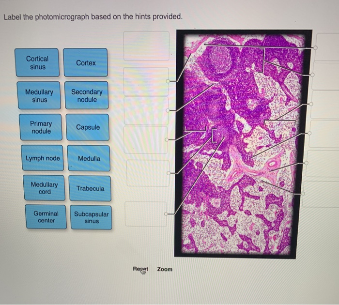
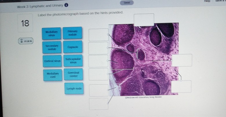
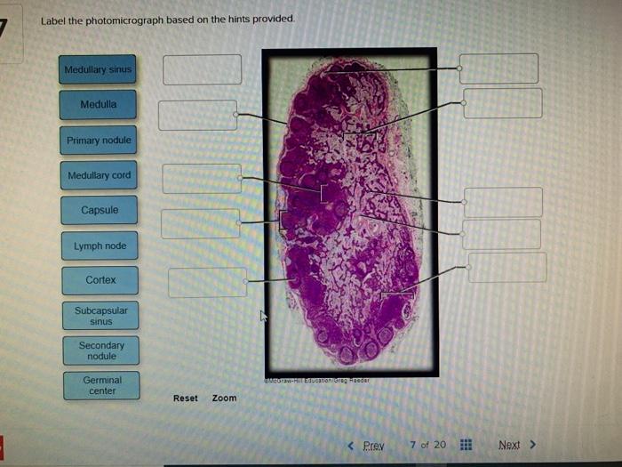
Capsule Capsule Subcapsular sinus Mantle zone Lymph node Lymph node Germinal center Germinal center Subcapsular sinus Mantle zone Reticular fiber Reticular fiber. Label the photomicrograph using the hints provided. The values underneath 60 mm hg is generally considered with the.
Label the photomicrograph based on the hints provided. Label the photomicrograph based on the hints provided. 100 1 rating Yes.
Label the photomicrograph based on the hints provided. Label the structures in the photomicrograph based on the hints provided label the layers of the tissue from the stomach using the hints if available label the mucous membrane tissue from the stomach using the hints if provided. Label the structures in the photomicrograph based on the hints provided in a typical blood capillary bed the balance of hydrostatic and colloid osmotic pressures results in filtration occurring at the arterial end of the capillaries.
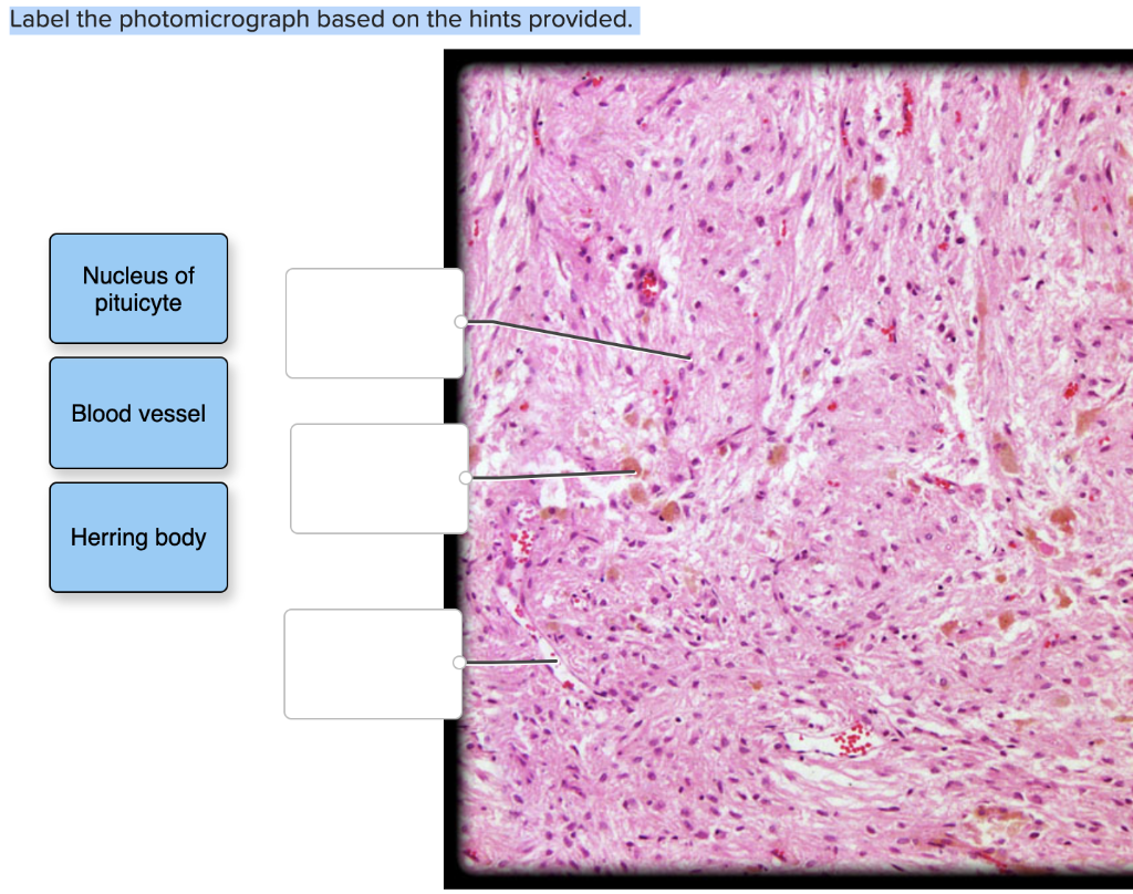
Distinguish the different types of pituitary cells using the. Its cells are pale staining and organized in ovoid clusters that are. Mantle zone Lymph node Subcapsular sinus Germinal center Capsule Mwl.
Label the structures in the photomicrograph based on the hints provided. Place each of the following lymphatic structures in the correct category based on their location. Label the structures in the photomicrograph based on the hints provided.
Label the photomicrograph based on the hints provided. Place each of the following lymphatic structures in the correct category based on their location. The adrenals are paired endocrine glands that sit anterosuperior and slightly.
Searchable by topic and provided in ms word format as well as in launchpad and diploma the assessment bank offers a high level of flexibility. Label the photomicrograph based on the hints provided. Label the photomicrograph based on the hints provided.
Place the following tonsils in order based on their location from superior to inferior. Correctly label the following anatomical parts of a kidney. Cortical sinus cortex medullary sinus secondary nodule primary nodule capsule lymph node medulla.
Chief cells appear as a dark purple in an HE stain with the oxyphil. Label the photomicrograph based on the hints provided.

Practical 2 Flashcards Quizlet

Endocrine Lab Flashcards Quizlet

Label The Structures In The Photomicrograph Based On The Hints Provided Spleen Capsule Capsule White Pulp Homeworklib

11 14 Test Flashcards Quizlet
Solved Label The Photomicrograph Based On The Hints Chegg Com
Solved Week 3 Lymphatic And Urinary Label The Chegg Com

Solved Label The Photomicrograph Based On The Hints Chegg Com

Endocrine System Apr Module 8 Flashcards Quizlet

Endocrine System Apr Module 8 Flashcards Quizlet
Solved Label The Photomicrograph Based On The Hints Chegg Com

Practical 2 Flashcards Quizlet

Endocrine System Apr Module 8 Flashcards Quizlet

Solved Label The Photomicrograph Based On The Hints Chegg Com
Solved Label The Photomicrograph Based On The Hints Chegg Com
Solved Label The Photomicrograph Based On The Hints Provided Chegg Com
Solved Please See An Attachment For Details Course Hero
Solved Lab 11 Endocrine System Saved Label The Chegg Com
Solved Label The Photomicrograph Based On The Hints Chegg Com
Solved Label The Photomicrograph Based On The Hints Chegg Com


Comments
Post a Comment